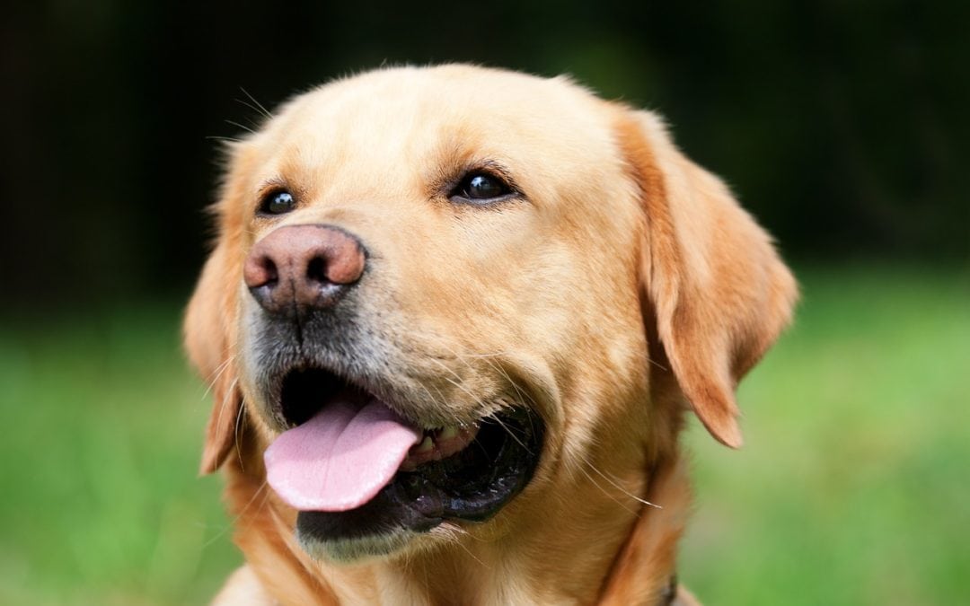
Sage came in on Monday afternoon. This two-year-old yellow lab had been hit by a car earlier in the day, but dad had to get home to help get her in the car. On first glance, she looked okay, but on closer inspection, she was a little shocky. Her gums were dry (a sign of dehydration) and her capillary refill time was about three seconds (a sign of shock).
But Sage was up on her feet and looking pretty good. At least she was on three feet and would touch the other one down. I palpated and did not feel a long bone fracture. So when choosing if I got to do either IV fluids or radiographs, I chose fluids.
IV fluids make everything better. They are the treatment of choice for shock, great for dehydration and even help with pain management. Ideally, we would do chest radiographs, abdominal radiographs, blood work and IV fluids, but often the economic reality does not allow for academically determined best care. IV fluids make the animal feel and do better, so they are always my first choice, but chest rads are a close second.
When an animal is hit by a car, it picks up the momentum of the car. Then when it hits the ground, it comes to an abrupt halt. This sudden stop can twist the animal inside its body. Often this shows up as a torn lung or pneumothorax. This is why chest rads are important. A small hole will seel and then heal. A large hole may leak enough air to keep the animal from being able to inflate their lungs due to the pressure outside the lungs. Without inflation, there is no air exchange and they die. But if a pneumothorax is found on the radiographs (x-rays), it can be treated. This is why rads are so important.
Abdominal radiographs are important for the same reason. Livers, spleens, kidneys, bladders, and intestines can all tear, but since there is more space and mobility in the abdomen than the chest, it is less likely. While I am fairly pushy about chest rads, I do not advocate for Abdominal rads as much.
Sage didn’t get either set of radiographs the first night and that is okay. I usually say that for an animal to heal, it is one-third me (I have to know what to do and do it), one-third the owners and one-third the pet. Sometimes owners cannot afford everything either emotionally or economically. Sometimes the animal has to do part of the healing on their own. If Sage had a pneumothorax and shock and a fracture, it might have been more than her parents could afford. I knew we would have to reserve something to take care of the unused leg, but hoped it would only be pain medicine for a fractured pelvis.
Tuesday morning, Sage looked a lot better! The fluids had done their job and her tail was wagging and she was bright and alert. But her leg was swollen more than the night before. I recommended that we do radiographs to evaluate the damage. Radiographs confirmed that she had a dislocated hip in addition to a mildly fractured pelvis. The pelvis is the box of bones that attack the spine to the back legs. It is a relatively common fracture but rarely requires surgery because the muscles around it support it during healing. The dislocated hip was going to require a bit of work.
Technically it is a coxofemoral luxation. The head of the femur is moved from the acetabular socket. Dislocation disrupts the joint capsule and other supportive structures of the hip, including ligaments and often bone. It is commonly caused by trauma, but joint degeneration or hip dysplasia can increase the risk. It is painful which is why she was not walking on it. Unlike a human shoulder luxation, it requires anesthesia and quite a lot of effort to reduce the luxation.
This is one of the few cases that I always get the textbook out for instructions. For cranial/dorsal luxations, first you externally rotate the leg, then significant traction and internal rotation followed by abducting the leg and flexing the knee and hock while simultaneously “guiding” the greater trochanter to drive the femoral head back into the socket. Now in my book that only takes about three sentences, but in real life that took over two hours with three of us working hard to move the leg into place. Of course, the book doesn’t mention hip dysplasia, torn ligaments, and muscles or other remnants of the trauma. It does say that only 50% can be reduced and stay in place. It merely brushes on the fact that radiographs may need to be taken intraoperatively to see where the leg is at that point. The problem is that the hip will pop over the rim of the acetabulum just like it does into the joint. It will move in almost normal fashion above the pelvis. Too much forceful movement to evaluate the position and the hip will have the cartilage damaged. After one of the radiographs to take stock of the position, the luxated hip was in a ventral position to the pelvis. The book said to do a combination of lifting, torquing and pushing. While I was trying to get a feel for the mechanics, but allowing Stephanie and Becky to rest a moment, the leg slid right into place. They perked up in disbelief, but we rotated the hip under pressure to move out any clots and it seemed stable.
Radiographs confirmed that the hip was back in place and the book was moved to the instructions for an Ehmer sling. This is a sticky stretchy figure of eight combination bandage that forces the hip into the socket while the muscles heal, but if the first rotation is not started in the correct manner, it doesn’t work. Hence the need for the textbook again.
With pain meds, Sage woke up wagging her tail. I am certain that she will get tired of the bandage before her “several weeks” of resting the leg are up. But Thursday evening, Sage went home. Wagging her tail and hopping on three legs, she was a tad brighter than when she came in.
Dr. MJ Wixsom owns and practices at Guardian Animal Medical Center on Bellefonte Road in Flatwoods, KY. 606.928.6566 and online at www.GuardianAnimal.com and has her fourth book out.